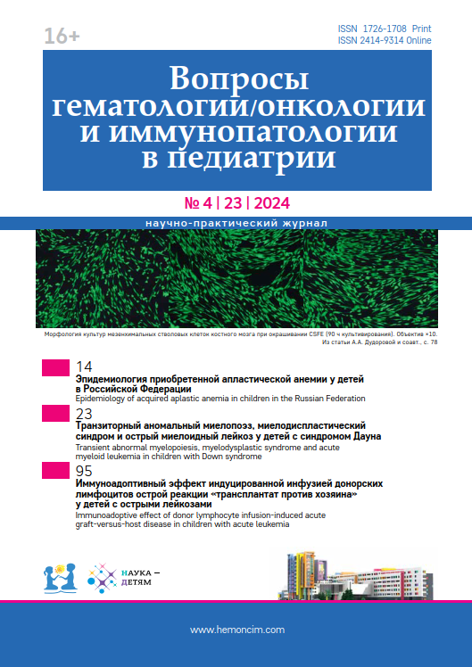
Identification of optimal conditions for the expansion of human bone marrow-derived mesenchymal stem cells using tools for live-cell culture monitoring
- Authors: Dudorova A.A.1, Efimenko M.V.1, Khismatullina R.D.1, Maschan M.A.1, Kazmina I.N.1, Ilyushina M.A.1, Osipova E.Y.1
-
Affiliations:
- The Dmitry Rogachev National Medical Research Center of Pediatric Hematology, Oncology and Immunology of Ministry of Healthcare of the Russian Federation
- Issue: Vol 23, No 4 (2024)
- Pages: 78-83
- Section: ORIGINAL ARTICLES
- Submitted: 12.02.2024
- Accepted: 08.04.2024
- Published: 13.12.2024
- URL: https://hemoncim.com/jour/article/view/820
- DOI: https://doi.org/10.24287/1726-1708-2024-23-4-78-83
- ID: 820
Cite item
Full Text
Abstract
Based on the results of our study, we have developed recommendations regarding cell culture media composition for the expansion of human bone marrow-derived mesenchymal stem cells (MSC) for preclinical studies and potential clinical applications. ALPHA-MEM supplemented with 10% platelet lysate proved to be the most effective culture medium. Different DMEM media supplemented with fetal bovine serum turned out to be less effective: a maximum of 80% confluence was reached after 80 hours of culture, while MSC confluence in StemMACS and ALPHA-MEM media supplemented with platelet lysate kept increasing even after 100 hours of expansion. The growth rate of MSCs in RPMI-1640 medium was significantly lower than in the other culture media. When culturing MSCs in media with high glucose concentration (4.5 g/L), the percentage of cells with fat transformation after 5 days of culture was higher than in low-glucose (1.0 g/L) media such as DMEM low gl, StemMacs, ALPHAMEM. It is preferable to use MSC expansion media that do not induce spontaneous adipogenic differentiation for culturing MSCs for clinical purposes because the cells remain uncommitted and all their differentiation potential can be used in accordance with the objectives of further research and/or clinical needs. This study was supported by the local Ethics Committee and approved by the Scientific Council of the Dmitry Rogachev National Medical Research Center of Pediatric Hematology, Oncology and Immunology of Ministry of Healthcare of the Russian Federation. All the participants signed the standard informed consent form and agreed to the use of some of their biological materials for research purposes.
About the authors
A. A. Dudorova
The Dmitry Rogachev National Medical Research Center of Pediatric Hematology, Oncology and Immunology of Ministry of Healthcare of the Russian Federation
Email: dudorova_aa@mail.ru
ORCID iD: 0000-0001-9444-4689
Moscow
Russian FederationM. V. Efimenko
The Dmitry Rogachev National Medical Research Center of Pediatric Hematology, Oncology and Immunology of Ministry of Healthcare of the Russian Federation
Email: masik_007@mail.ru
ORCID iD: 0000-0002-3001-4820
Moscow
Russian FederationR. D. Khismatullina
The Dmitry Rogachev National Medical Research Center of Pediatric Hematology, Oncology and Immunology of Ministry of Healthcare of the Russian Federation
Email: Rimma.Chismatullina@fccho-moscow.ru
ORCID iD: 0000-0001-5618-7159
Moscow
Russian FederationM. A. Maschan
The Dmitry Rogachev National Medical Research Center of Pediatric Hematology, Oncology and Immunology of Ministry of Healthcare of the Russian Federation
Email: mmaschan@yandex.ru
ORCID iD: 0000-0003-1735-0093
Moscow
Russian FederationI. N. Kazmina
The Dmitry Rogachev National Medical Research Center of Pediatric Hematology, Oncology and Immunology of Ministry of Healthcare of the Russian Federation
Email: rinakazmina@mail.ru
ORCID iD: 0009-0000-0448-504X
Moscow
Russian FederationM. A. Ilyushina
The Dmitry Rogachev National Medical Research Center of Pediatric Hematology, Oncology and Immunology of Ministry of Healthcare of the Russian Federation
Email: Mariya.Ilyushina@fccho-moscow.ru
ORCID iD: 0000-0001-7652-7704
Moscow
Russian FederationE. Yu. Osipova
The Dmitry Rogachev National Medical Research Center of Pediatric Hematology, Oncology and Immunology of Ministry of Healthcare of the Russian Federation
Author for correspondence.
Email: e_ossipova@mail.ru
ORCID iD: 0000-0002-1873-3486
Elena Yu. Osipova, Head of the Laboratory of Physiology and Pathology of Stem Cells
1 Samory Mashela St., Moscow 117997
Russian FederationReferences
- Frieedenstein A.J., Petrakova K.V., Kurolesova A.I., Frolova G.P. Heterotopic of bone marrow. Analysis of precursor cells for osteogenic and hematopoietic tissues. Transplantation 1968; 6 (2): 230–47.
- Kulus M., Sibiak R., Stefanska K., Zdun M., Wieczorkiewicz M., Piotrowska-Kempisty H., et al. Mesenchymal Stem/Stromal Cells Derived from Human and Animal Perinatal Tissues – Origins, Characteristics, Signaling Pathways, and Clinical Trials. Cells 2021; 10 (12): 3278.
- Lazarus H.M., Haynesworth S.E., Gerson S.L., Rosenthal N.S., Caplan A.I. Ex vivo expansion and subsequent infusion of human bone marrow-derived stromal progenitor cells (mesenchymal progenitor cells): implications for therapeutic use. Bone Marrow Transplant 1995; 16 (4): 557–64.
- Golpanian S., Wolf A., Hatzistergos K., Hare J.M. Rebuilding the damaged heart: mesenchymal stem cells, cell-based therapy, and engineered heart tissue. Physiol Rev 2016; 96 (3): 1127–68.
- Hoang D.M., Pham P.T., Bach T.Q., Ngo A.T., Nguyen Q.T., Phan T.T., et al. Stem cell-based therapy for human diseases. Signal Transduct Target Ther 2022; 7 (1): 1–41.
- Zhou L., Zhu H., Bai X., Huang J., Chen Y., Wen J., et al. Potential mechanisms and therapeutic targets of mesenchymal stem cell transplantation for ischemic stroke. Stem Cell Res Ther 2022; 13 (1): 195–210.
- Han H.T., Jin W.L., Li X. Mesenchymal stem cells-based therapy in liver diseases. Mol Biomed 2022; 3 (1): 23–74.
- Gavin C., Boberg E., Von Bahr L., Bottai M., Andrén A.T., Wernerson A., et al. Tissue immune profiles supporting response to mesenchymal stromal cell therapy in acute graft-versus-host disease – a gut feeling. Stem Cell Res Ther 2019; 10: 334– 40.
- Dabrowska S., Andrzejewska A., Janowski M., Lukomska B. Immunomodulatory and regenerative effects of mesenchymal stem cells and extracellular vesicles: therapeutic outlook for inflammatory and degenerative diseases. Front Immunol 2021; 11: 1–26.
- Ciervo Y., Ning K., Jun X., Shaw P.J., Mead R.J. Advances, challenges and future directions for stem cell therapy in amyotrophic lateral sclerosis. Mol Neurodegener 2017; 12 (1): 85–107.
- Leyendecker J.A., Gomes Pinheiro C.C., Amano M.T., Franco Bueno D. The use of human mesenchymal stem cells as therapeutic agents for the in vivo treatment of immune-related diseases: a systematic review. Front Immunol 2018; 9: 1–51.
- Re F., Borsani E., Rezzani R., Sartore L., Russo D. Bone regeneration using mesenchymal stromal cells and biocompatible scaffolds: A concise rewiew of the current clinical trials. Gels 2023; 9 (5): 389–404.
- Nikolaeva Yu.A., Skorobogatova E.V., Kirgizov K.I., Pristanskova E.A., Blagonravova O.L., Osipova E.U., et al. Intraosseous infusion of mesenchymal stem cells in treatment of graft-versus-host refractory response with graft hypofunction. Pediatrics. Journal named after G.N. Speransky 2019; 98 (4): 8–14. (In Russ.)
- Dominici M., Le Blanc K., Mueller I., Slaper-Cortenbach I., Marini F.C., Krause D.S., et al. Minimal criteria for defining multipotent mesenchymal stromal cells. The International Society for Cellular Therapy position statement. Cytotherapy 2006; 8 (4): 315–7.
- Mushahary D., Spittler A., Kasper C., Weber V., Charwat V. Isolation, cultivation and characterization of human mesenchymal stem cells. Cytometry A 2018; 93 (1): 19–31.
- Viau S., Lagrange A., Chabrand L., Lorant J., Charrier M., Rouger K., et al. A highly standardized and characterized human platelet lysate for efficient and reproducible expansion of human bone marrow mesenchymal stromal cells. Cytotherapy 2019; 21 (7): 738–54.
- Shamanskaya T.V., Osipova E.Yu., Roumiantsev S.A. Mesenchymal stem cells ex vivo cultivation technologies for clinical use. Oncohematology 2009; (3): 69–76. (In Russ.) doi: 10.17650/1818-8346-2009-0-3-69-76
- Astakhova N.M., Korel A.V., Kirilova I.A. The development of protocols for isolation, culture and differentiation of human bone marrow-derived mesenchymal stem cells. Genes and Cells 2017; 12 (3): 128. (In Russ.)
- Wong C.W., Han H.W., Hsu S.H. Changes of cell membrane fluidity for mesenchymal stem cell spheroid on biomaterial surfaces. World J Stem Cells 2022; 14 (8): 616–32.
- Jakl V., Popp T., Haupt J., Port M., Roesler R., Wiese S. Effect of expansion Media on functional characteristics of bone marrow-derived mesenchymal stromal cells. Cells 2023; 12 (16): 2105–36.
- Toth G., Szollosi J., Vereb G. Quantitating ADCC against adherent cells: impedance based detection is superior to release, membrane permeability, or caspase activation assays in resolving antibody dose response. Cytometry A 2017; 91 (10): 1021–9.
- He L., Li M., Wang X., Wu X., Yue G., Wang T., et al. Morphology-based deep learning enables accurate detection of senescence in mesenchymal stem cell cultures. BMC Biol 2024; 22 (1): 1–17.
- Parvin Nejad S., Lecce M., Mirani B., Machado Siqueira N., Mirzaei Z., Santerre J.P., et al. Serum- and xeno-free culture of human umbilical cord perivascular cells for pediatric heart valve tissue engineering. Stem Cell Res Ther 2023; 14 (1): 96–111.
- Even K.M., Gaesser A.M., Ciamillo S.A., Linardi R.L., Ortved K.F. Comparing the immunomodulatory properties of equine BM-MSC culture expanded in autologous platelet lysate, pooled platelet lysate, equine serum and fetal bovine serum supplemented culture media. Front Vet Sci 2022; 25 (9): 1–14.
- Fitzgerald J.C., Shaw G., Murphy J.M., Barry F. Media matters: culture medium-dependent hypervariable phenotype of mesemchymal stromal cells. Stem Cell Res Ther 2023; 14 (1): 363–78.
- Altrock E., Sens-Albert C., Hofmann F., Riabov V., Schmitt N., Xu Q., et al. Significant improvements of bone marrow-derived MSC expansion fron MDS patients by defined xeno-free medium. Stem Cell Res Ther 2023; 14 (1): 156–64.
Supplementary files


