


Pediatric Hematology/Oncology and Immunopathology
Peer-review quarterly medical journal
Editor-in-Chief
- Galina A. Novichkova, Doctor of Medical Sciences, Professor
ORCID: 0000-0003-4911-0553
Founders
- Dmitry Rogachev National Medical Research Center of Pediatric Hematology, Oncology and Immunology
WEB: https://fnkc.ru/ - Foundation for Support and Development in the Field of Paediatric Haematology, Oncology and Immunology "Science to Children"
WEB: https://fnkc.ru/vind-fnkc/
Publisher
- LLC «Science and education»
About
The main aspects of the articles published in the journal are Clinical Medicine, Pediatrics and Children's Health, Hematology, Oncology, Immunology and Allergic Reactions, Organization of Specialized Care for Children. The journal features research findings on the efficacy and safety of new medications, discusses current treatment protocols for oncological, hematological and immunological diseases, analyses international experience concerning the use of various drugs and diagnostic methods.
The Editorial Board and Advisory Editors include leading experts of the Center as well as our counterparts from Austria, Germany, Israel, the Netherlands, the USA, France and Japan. Thanks to our long-lasting and effective cooperation, important steps have been taken to develop and implement modern medical advances into healthcare in Russia.
The journal is indexed by Scopus, Ulrich’s Periodicals Directory and Russian Science Citation Index. The journal is included in the List of leading peer-reviewed scientific journals and periodicals of Higher Attestation Commission of the Ministry of Education of Russia, where key results of doctoral theses should be published.
Standard journal size: А4; number of pages: 96. Journal circulation – 3000. The journal is distributed by subscription via Rospechat Agency and among the participants of events arranged by D. Rogachev NMRCPHOI and regional public organization “National Society of Pediatric Hematologists and Oncologists”. Advertisement placement: 2nd, 3rd and 4th cover pages, a two-page spread and an insert. Advertisement size: full А4 page, half page, one-third page and quarter page.
Current Issue
Vol 24, No 2 (2025)
- Year: 2025
- Published: 30.06.2025
- Articles: 17
- URL: https://hemoncim.com/jour/issue/view/58
-
Description:
Published - 30.06.2025
Full Issue

ORIGINAL ARTICLES
Preclinical development of a protocol for the manufacturing of anti-CD5 CAR-T lymphocytes for the treatment of T-lineage acute lymphoblastic leukemia and lymphoma
Abstract
The development of cell therapy methods for T-lineage acute lymphoblastic leukemia remains a relevant clinical challenge. The current study presents a preclinical stage of anti-CD5 CAR-T cell development without the use of additional genetic modifications, relying on down-regulation of CD5. The resulting cell products demonstrated sufficient expansion rates and CAR expression levels, the predominance of the Tem phenotype (CD197–CD45RA–) and low expression of exhaustion markers (PD-1, CD57, TIGIT). The antitumor function was confirmed both in vitro through the assessment of degranulation, cytokine production, and cytotoxicity against the Jurkat cell line, and in vivo through the evaluation of CD5+ leukemia elimination in a murine model. The data obtained may serve as a basis for the development of a clinical manufacturing protocol for anti-CD5 CAR-T cells. The study was approved by the Independent Ethics Committee and the Scientific Council of the Dmitry Rogachev National Medical Research Center of Pediatric Hematology, Oncology and Immunology.
 14-26
14-26


Experience in developing and scaling a protocol for NK lymphocyte enriched cellular product manufacturing for clinical application
Abstract
for NK lymphocyte-enriched biomedical cell product is a pressing issue in clinical biotechnology. Based on numerous scientific and clinical studies, we have settled on the NK lymphocyte expansion protocol with a modified feeder line K562 and peripheral
blood mononuclear cells as a starting fraction, not including CD3+ lymphocyte depletion step. In this paper, we present the results of a validation process of NK lymphocyte-enriched biomedical cell product manufacturing. The study was approved by the Independent Ethics Committee and the Scientific Council of the Dmitry Rogachev National Medical Research Center of Pediatric Hematology, Oncology and Immunology of Ministry of Healthcare of the Russian Federation. The resulting cell products
met all stated quality control criteria, contained a high percentage of NK cells, and demonstrated strong cytotoxic activity against the THP-1 and Jeko-I cell lines. Expansion folds allowed us to achieve the desired clinical dosage in all processes. The
surface marker expression profile of the expanded NK cells corresponded to an activated and highly proliferative phenotype. The residual lymphocytes were represented mainly by T cells with an effector phenotype, and a significant decrease in TCRab percentage was observed compared to the initial fraction. These data provide the basis for a clinical study of the feasibility and safety of the infusions of the cellular products produced according to this protocol for pediatric refractory leukemia treatment.
27-37
Post-transplant cyclophosphamide, abatacept, and vedolizumab for the prevention of graft-versus-host disease after hematopoietic stem cell transplantation in children with acute leukemia: results of a prospective study
Abstract
High-dose post-transplant cyclophosphamide (PTCy) is an established method for the prevention of graft-versus-host disease (GVHD) in allogeneic hematopoietic stem cell transplantation (HSCT) from haploidentical donors, however data on its efficacy in the pediatric population remain limited. In this prospective study, we evaluated an improved PTCy-based GVHD prophylaxis regimen comprising the a4b7 integrin blocker vedolizumab and a co-stimulatory signal inhibitor abatacept in addition to standard treatment with PTCy and cyclosporine A. The study was approved by the Independent Ethics Committee and the Scientific Council of the Dmitry Rogachev National Medical Research Center of Pediatric Hematology, Oncology and Immunology of Ministry of Healthcare of the Russian Federation. Allogeneic HSCT was performed in children with high-risk acute leukemia in complete remission from a haploidentical (n = 54) or unrelated (n = 4) donor. Myeloablative conditioning was based on treosulfan or total body irradiation. In 97% of cases, bone marrow was used as a graft source. The engraftment rate was 98%. The cumulative risk of acute GVHD grade II-IV and III-IV was 38% and 8.6%, respectively, of acute intestinal GVHD grade II-IV - 10%, and of chronic GVHD - 7%. Transplant-related mortality after 2 years of follow-up was 3.6%, the incidence of relapse was 16%, the overall survival rate - 91%, and the event-free survival rate - 85%. Overall, the proposed GVHD prophylaxis regimen demonstrated a favorable safety profile and good tolerability, no specific adverse events were observed.
38-45
Gonadal function in male pediatric patients after allogeneic hematopoietic stem cell transplantation: a comparison of the effects of treosulfan- and total body irradiation-based conditioning
Abstract
Allogeneic hematopoietic stem cell transplantation (HSCT) is the only curative treatment for many orphan diseases that can significantly improve prognosis and survival in patients. Conditioning prior to HSCT includes alkylating agents and/or total body irradiation (TBI) that are both known to be highly gonadotoxic. This is why it is so important to choose drugs and treatment options with minimal side effects. The aim of our study was to evaluate the reproductive function of male HSCT recipients who had received treosulfan- and TBI-based conditioning. The study was approved by the Independent Ethics Committee and the Scientific Council of the Dmitry Rogachev National Medical Research Center of Pediatric Hematology, Oncology and Immunology of Ministry of Healthcare of the Russian Federation. We included 112 boys. The concentrations of follicle-stimulating and luteinizing hormones, testosterone levels, testicular size (measured using ultrasound) and pubertal development (as per the Tanner scale) were assessed over time. The obtained data showed that the effect of treosulfan on gonadal function in the males was less significant than that of TBI, with the cumulative probability of developing hypogonadism at 4 years after HSCT equaling 0% and 24% respectively. However, the toxicity of other alkylating agents included in a treosulfan-based conditioning regimen eliminates the difference in the cumulative probability of hypogonadism between the two approaches. Further annual monitoring is needed to fully understand the reproductive potential of the patients.
46-54
Outcomes of hematopoietic stem cell transplantation using melphalan-based conditioning in children with acquired aplastic anemia
Abstract
Despite the high effectiveness of hematopoietic stem cell transplantation (HSCT) in children with acquired aplastic anemia (AAA), the procedure can still result in such major issues as graft failure and rejection. Risk factors for graft rejection and poor function as well as the persistence of mixed chimerism include allosensitization, insufficient number of transplanted cells and relatively preserved native myelopoiesis. Long-term engraftment can be achieved by modifying conditioning and immunosuppressive therapy. One such promising approach is the inclusion of melphalan in a conditioning regimen. In our study, we retrospectively analyzed outcomes of HSCT with melphalan-based conditioning carried out in 21 children with AAA (for 18 patients, it was their first HSCT, while 3 had a repeat HSCT): out of these, 11 patients received transplants from matched unrelated donors, 9 received transplants from matched related donors and 1 - from a haploidentical donor. Nine children received calcineurin inhibitor-based graft-versus-host disease prophylaxis while 12 children received Janus kinase inhibitors. A good graft function was achieved in all the children, without any severe complications. As per the latest findings, 95% of the patients achieved full total donor cell chimerism, 85.7% of the patients had full donor chimerism in the CD3+ cell lineage, while 14.3% had mixed CD3+ cell chimerism with 9-13% of own cells. The inclusion of melphalan in a conditioning regimen in patients with AAA before HSCT in combination with new less toxic graft-versus-host disease prophylaxis regimens may be an effective strategy to overcome the risk of graft rejection and ensure a low toxicity profile and low incidence of complications. The study was approved by the Independent Ethics Committee and the Scientific Council of the N.I. Pirogov Russian National Research Medical University of Ministry of Healthcare of the Russian Federation.
55-61
Results of allogeneic hematopoietic stem cell transplantation from a genoidentical sibling after high-dose treosulfan-based conditioning in children with intermediate-risk acute myeloid leukemia in first clinical and hematological remission
Abstract
The value of allogeneic hematopoietic stem cell transplantation (HSCT) in first remission in children with intermediate-risk acute myeloid leukemia (AML) remains the matter of debate. We analyze the outcome of HSCT from a genoidentical sibling with high-dose treosulfan-based myeloablative conditioning and enhanced graft-versus-host disease (GVHD) prophylaxis in comparison with continuation of high-dose chemotherapy (CT). The study was approved by the Independent Ethics Committee and the Scientific Council of the Dmitry Rogachev National Medical Research Center of Pediatric Hematology, Oncology and Immunology of Ministry of Healthcare of the Russian Federation. A group of interest consisted of 22 HSCT recipients and a comparison group consisted of 240 patients who received high-dose CT. The conditioning regimens included treosulfan (n = 15), busulfan (n = 3), and melphalan (n = 4). The graft source was bone marrow in 12 patients and peripheral stem cells in 10 patients. GVHD prophylaxis was intensified either by including 1 or 2 additional immunosuppressive drugs or by increasing the duration of prophylaxis. The comparison group consisted of 240 patients who received 1 or 2 cycles of high-dose cytarabine- based consolidation therapy. The groups were matched for all initial characteristics. Engraftment was achieved in all 22 HSCT recipients: the median time to granulocyte recovery was day +16 (range, day +11 - day +21) and the median time to platelet recovery was day +15 (range, day +11 - day +28). The main toxicities of the conditioning regimens were mucositis in 12 (54%) patients, skin lesions in 6 (27%) patients, and an increase of alanine aminotransferase in 4 (18%) patients. There were no cases of toxicity-related mortality. Acute and chronic GVHD, requiring medical treatment developed in 15% of the patients. Four HSCT recipients relapsed between 3 months to 3 years after HSCT. In the high-dose CT group, 24 (10%) patients died of infectious complications and 79 (37%) patients developed a relapse. The cumulative incidence of relapse was 0.23 ± 0.10 and 0.44 ± 0.4 and the probability of the overall survival was 0.88 ± 0.08 and 0.83 ± 0.03 in the HSCT group and high-dose CT group, respectively (p > 0.05). HSCT from a genoidentical sibling in intermediate-risk AML patients in first clinical and hematological remission is a safe and effective procedure, and tends to reduce the risk of AML relapse.
62-72
Hereditary factor XIII deficiency in children: diagnostic and clinical features and clinical experience with replacement therapy
Abstract
Factor XIII deficiency is an extremely rare bleeding disorder with a prevalence of 1 in 2–3 millions. This deficiency is characterized by severe bleeding manifestations: life-threatening bleeding, including recurrent bleeding into the central nervous system. Here, we report 16 cases of factor XIII deficiency in children who were under our observation at the beginning of 2025. The use of factor XIII concentrate is a global standard for the treatment of patients with factor XIII deficiency. Since 2023, children with confirmed hereditary factor XIII deficiency have been receiving this concentrate with the help of the Circle of Kindness Foundation. By the time of publication, 9 out of 12 children who had been prescribed treatment with factor XIII concentrate started to receive it. All the patients receiving this therapy do not have spontaneous bleeding. In this article, we describe clinical and phenotypical features as well as bleeding manifestations in the patients with factor XIII deficiency and present our experience with factor XIII concentrate therapy. Ethical approval was not required since the study involved the use of anonymized retrospective data obtained during routine clinical practice.
74-79
Cytofluorometry analysis of red blood cell suspension properties: results of a pilot study
Abstract
A wide use of blood products inevitably leads to an increase in the number of complications associated with it. These complications include thromboembolic events that are considered to be rare and difficult-to-manage. Currently, the mechanisms and possible laboratory predictors of such complications are unclear. This paper presents our experience of using flow cytometry to assess the parameters of phosphatidylserine expression (detected by Annexin V binding) and describes impairments in the deformability of red blood cells (detected using forward and side scatter data) which determine the procoagulant properties of cells. According to our findings, the use of Annexin V for the assessment of gradual cell death in the samples of red blood cell (RBC) suspension is controversial. At the same time, our results show a higher sensitivity of the assessment of the accumulation of RBCs with an altered shape for the detection of programmed cell death - eryptosis. The obtained results can be used to improve the detection of dead RBCs during the routine storage of RBC suspension. The study was approved by the Independent Ethics Committee and the Scientific Council of the Dmitry Rogachev National Medical Research Center of Pediatric Hematology, Oncology and Immunology of Ministry of Healthcare of the Russian Federation.
80-85
The effect of two red-cell transfusion strategies on outcomes and incidence of anemia after discharge in premature infants with extremely low birth weight
Abstract
Premature newborns with extremely low birth weight are the main category of infants having to undergo one or more red-cell transfusions during the neonatal period in order to treat anemia. Presently, there are two major transfusion strategies: the liberal strategy aimed at maintaining higher hemoglobin levels, and the restrictive one when blood is transfused at lower hemoglobin levels. In our study, we aimed to compare outcomes and the incidence of anemia after discharge during the first 24 months of corrected age in infants with extremely low birth weight who had received red-cell transfusions according to the restrictive or liberal strategy. The study was approved by the Biomedical Research Ethics Committee (minutes No.12 of 17 November 2016) and the Scientific Council (minutes No.19 of 29 November 2016) of the V.I. Kulakov National Medical Research Center for Obstetrics, Gynecology and Perinatology of Ministry of Healthcare of Russia. This was a single-center retrospective cohort study carried out at clinical departments of the Institute of Neonatology and Pediatrics of the V.I. Kulakov National Medical Research Center for Obstetrics, Gynecology and Perinatology of Ministry of Healthcare of Russia. The included infants were divided into 2 groups: group 1 – infants who had received RBC transfusions following the liberal strategy; group 2 – infants in whom the restrictive transfusion strategy had been used. We looked at the incidence of such outcomes as retinopathy of prematurity (ROP) ≥ stage III, necrotizing enterocolitis (NEC) ≥ stage II, bronchopulmonary dysplasia (BPD), intraventricular hemorrhages (IVH) ≥ grade II, periventricular leukomalacia (PVL), anemia after discharge at a corrected age (CA) of 1, 3, 6, 12, 18 and 24 months. The gestational age of the newborns ranged from 24 to 28 weeks. The frequency of red-cell transfusions was 136/164 (82.9%) in group 1 versus 149/216 (68.9%) in group 2, with the median volume of transfusions of 29.3 mL/kg (IQR 17.5–51.8 mL/kg) in group 1 and 19.5 mL/kg (IQR 15.0–29.5 mL/kg) in group 2, p < 0.05. There were no significant differences in the incidence of BPD, IVH ≥ grade II, PVL, ROP ≥ stage III between the study groups. The incidence of NEC ≥ stage II was 1.6 times higher in group 2 than in group 1. The incidence of anemia after discharge was lower in group 1 compared to group 2 at a CA of 1 and 3 months (32/72 (44.4%) vs. 106/166 (63.8%), p = 0.026 at 1 month, 28/72 (38.9%) vs. 90/166 (54.2%), p = 0.042 at 3 months). There were no reports of severe anemia after discharge in either of the groups. After 6 months CA there was no difference in the incidence of anemia between the groups. The use of the restrictive red-cell transfusion strategy did not lead to a clinically significant increase in the incidence of ROP ≥ stage III, BPD, PVL, IVH ≥ grade II or anemia after 6 months of CA. However, the risk of NEC ≥ stage II in the restrictive strategy group was 1.6 times higher than in the liberal one, which requires further analysis.
86-94
Morphology of a pure hematogone population isolated from normal and hematogone-rich bone marrow
Abstract
B-ceLL precursors (hematogones) account for Less than 5% of normaL bone marrow (BM) nucLeated ceUs but in some cLinicaL conditions the number of hematogones can be transientLy increased. Their morphoLogy has been characterized in patients with increased numbers of hematogones. However, the scarcity of hematogones and inabiLity to obtain their pure popuLation make it difficuLt to thoroughLy investigate the morphoLogy of these ceLLs in normaL BM. A pure popuLation of hematogones was isoLated as a fraction of CD10+ BM mononucLear ceLLs from 14 patients with increased numbers of hematogones and 12 heaLthy donors using microarrays with immobiLized antibodies to duster of differentiation (CD) antigens. We described the main morphoLogicaL subtypes of hematogones of normaL BM and anaLyzed the proportion of these ceUs among CD19+, CD22+, CD20+, and CD10+CD34+ BM mononucLear ceUs. One to nine percent of hematogones are 13-17 mm in diameter and have basophiLic cytopLasm, Lacy or fineLy reticuLated, bLastic chromatin and prominent nucLeoLi. The remaining hematogones are immature Lymphoid ceLLs that vary considerabLy in size (7-15 mm in diameter) and have scanty cytopLasm, smudged chromatin and absent nucLeoLi. Three to twenty-five percent of these ceLLs are Larger than 10 mm. CD10+CD34+ ceLLs do not show predominancy bLastic morphoLogy, however some bLast-Like hematogones express CD20. Hematogones in normaL BM are heterogeneous in morphoLogy and size. We found no apparent correLation between the morphoLogy of hematogones and their stages of maturation. The study was approved by the Independent Ethics Committee and the Scientific CounciL of the Dmitry Rogachev NationaL MedicaL Research Center of Pediatric HematoLogy, OncoLogy and ImmunoLogy of Ministry of HeaLthcare of the Russian Federation. ALL donors and patients gave their informed consent to BM aspiration and participation in future biomedicaL studies.
96-103
Detection of phosphatidylserine-expressing red blood cells in whole blood
Abstract
There are various mechanisms of red blood cell (RBC) clearance from the circulation, including eryptosis and hemolysis. The spleen plays a major role in eliminating RBCs from the blood flow thanks to their ability to pass through narrow spaces. Still, some questions such as the role of phosphatidylserine-binding proteins in this process remain unanswered. Thus, RBC elimination mechanisms are still not very well explored. Here, we report on a flow cytometry protocol using phosphatidylserine markers for the detection of whole blood erythrocytes that can be classified as pre-elimination RBCs. We also provide the results of healthy donors' blood testing suggesting that reliable detection of eryptosis in whole blood is indeed possible. The study was approved by the Independent Ethics Committee and the Scientific Council of the Dmitry Rogachev National Medical Research Center of Pediatric Hematology, Oncology and Immunology of Ministry of Healthcare of Russia.
104-110
CLINICAL OBSERVATIONS
Severe hypercholesterolemia due to the cholestatic form of acute graft-versus-host disease with liver damage in a child following allogeneic hematopoietic stem cell transplantation
Abstract
Hypercholesterolemia is one of the complications of allogeneic hematopoietic stem cell transplantation. Various factors may contribute to its etiology, including graft-versus-host disease (GVHD) of the liver. This publication presents a clinical case of severe hypercholesterolemia associated with acute GVHD with the liver involvement in a patient with Nijmegen syndrome. The patient's parents gave their consent to the use of their child's data, including photographs, for research purposes and in publications. A high level of lipoprotein X was detected by peripheral blood electrophoresis and subsequent typing. This laboratory phenomenon is a characteristic manifestation of dyslipidaemia associated with cholestasis accompanying the hepatic form of GVHD. Clinical improvement and a reduction of cholesterol levels were achieved after intensification of immunosuppressive therapy. This clinical case highlights the necessity for early recognition and differential assessment of severe hypercholesterolemia in patients following allogeneic hematopoietic stem cell transplantation.
111-115
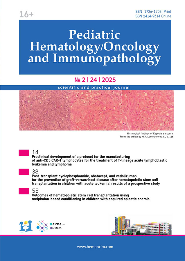
Disseminated Kaposi's sarcoma in a child treated with allogeneic hematopoietic stem cell transplantation
Abstract
Kaposi's sarcoma (KS) is a rare complication of hematopoietic stem cell transplantation (HSCT). Here, we report a case of disseminated KS in a pediatric patient after haploidentical HSCT that developed during secondary immunosuppression. Treatment with liposomal doxorubicin resulted in a complete metabolic response. This case highlights the importance of early diagnosis and stresses the need to consider KS in the differential diagnosis of cutaneous and systemic manifestations in patients following HSCT. The patient's parents gave consent to the use of their child's data, including photographs, for research purposes and in publications.
116-122
VISUALIZATION IN CLINICAL MEDICINE
A cutaneous manifestation of Stenotrophomonas maltophilia infection in a patient previously treated with allogeneic haematopoietic stem cell transplantation
Abstract
The article provides a brief description of a case of St. maltophilia metastatic skin lesion in a child diagnosed with primary immunodeficiency on the background of deep agranulocytosis associated with rejection of an allogeneic hematopoietic stem cell transplant.
123-123
LITERATURE REVIEW
Prolonged hypogammaglobulinemia in children after allogeneic hemopoietic stem cell transplantation
Abstract
Allogeneic hematopoietic stem cell transplantation (HSCT) is a key treatment strategy for life-threatening hematologic and immunologic diseases. Immune reconstitution after allogeneic HSCT is a complex and multistage process, which is critical to the recovery of the patient's immune function and to the reduction of the risk of infectious complications. Studies show that most patients experience a significant improvement in immune function 6-12 months after transplantation; however, a full recovery may take up to 2 years or more. One of the major complications of allogeneic HSCT is prolonged hypogammaglobulinemia - low serum immunoglobulin G levels for more than 2 years after allogeneic HSCT. In the absence of a clearly defined threshold for hypogammaglobulinemia, the generally accepted definition is a serum immunoglobulin G level below 5 g/L. Impaired antibody formation and the occurrence of prolonged hypogammaglobulinemia in children after allogeneic HSCT are complex problems that require careful monitoring and appropriate management. Timely restoration of B cell number and function is critical to reduce the risk of infectious complications and improve the quality of patients' life.
124-129
Congenital dyserythropoietic anemias
Abstract
Congenital dyserythropoietic anemias (CDAs) is a rare group of inherited anemias characterized by ineffective erythropoiesis and pronounced morphological abnormalities in erythroid precursors in the bone marrow. There are several CDA (I–IV) types, each associated with specific mutations in genes such as CDAN1, C15orf41, SEC23B, KIF23, and KLF1, leading to variable phenotypic manifestations. CDA type II is the most common variant among all CDAs and presents with normocytic anemia of varying severity, jaundice, and splenomegaly. The diagnosis of these genetically determined conditions is based on molecular genetic testing and morphological identification of specific abnormalities in erythroid precursors, with each type of CDA showing distinctive features, such as the presence of internuclear chromatin bridges in CDA type I and erythroblasts with two or more nuclei in CDA type II. Current treatment approaches play primarily a supportive role and include blood component transfusions, monitoring, and correction of iron overload. Hematopoietic stem cell transplantation is currently the only curative option for patients with CDA. Overall, the prognosis for this category of patients is favorable; however, the challenge lies in the heterogeneity of clinical and laboratory manifestations, which often makes it difficult to establish a definitive diagnosis.
130-139
New therapeutic options for the treatment of transfusion-dependent Diamond-Blackfan anemia
Abstract
Diamond-Blackfan anaemia is a rare congenital bone marrow failure syndrome characterized by suppression of erythropoiesis due to intense apoptosis of erythroid precursors resulting from defective ribosome biosynthesis. Classic options for the treatment of Diamond-Blackfan anaemia include long-term glucocorticosteroid therapy and transfusion of donor red blood cells. However, these approaches eventually lead to late adverse events, which stimulates the search for alternative therapies. In this paper, we review the current knowledge of the pathogenesis and therapy of this disease.
140-146